Biomaterials & Interface Phenomena
(BIOFIN)
Room JI204
About:
Biomaterials & Interface Implant-Tissues Phenomena is part of the Department of Metallic Materials Science, Physical Metallurgy, Faculty Materials Science and Engineering, University POLITEHNICA of Bucharest
The laboratory was established into the frame of the project Interdisciplinary Platform BIOENGINEERING-BIOTECHNOLOGY for Research, Development and Education – BIOINGTEH, funded by CNCSIS, 2006-2008, but some new equipment was added later from other research projects.
The research activities performed into the laboratory are integrated into Biomaterials Research Center –BIOMAT (national projects) and Research Department of University Politehnica of Bucharest (international projects).
Collaborations
National
- Technical University “Gh. Asachi” of Iassy
- Babes-Bolyai University Cluj-Napoca
- University Politehnica of Timisoara
- University Transilvania Brasov
- Medicine and Pharmacy University “Carol Davila” Bucharest
- Medicine and Pharmacy University “Gr. T. Popa” Iasi
- Medicine and Pharmacy University “V.Babes” Timisoara
- Medicine and Pharmacy University “Iuliu Hatieganu” Cluj-Napoca
- University “Titu Maiorescu” Bucharest
- University of Oradea
- University Ovidius Constanta
- Emergency University Clinical Hospital SUUB Bucharest
- Emergency Clinical Hospital Colentina Bucharest
- Emergency Clinical Hospital Floreasca Bucharest
- Emergency Clinical Hospital Elias Bucharest
- Central Universitary Emergency Military Hospital “Dr.Carol Davila” Bucharest
- Sanador Hospital Bucharest
- Institute of Cellular Biology and Pathology „Nicolae Simionescu” Bucharest
- Institute of Macromolecular Chemistry. "Petru Poni" Iasi.
- The National Institute for Laser, Plasma & Radiation Physics (INFLPR)
- INCDTP - Leather and Footwear Research Institute, Collagen Department
- Institute for Research in Chemistry “Raluca Ripan” Cluj-Napoca
- Medical Ortovit SRL Bucharest
- Vodimedicor SRL Bucharest
- Rodax Impex SRL Bucharest
- Ortopedica Bucharest
International
ERASMUS Agreements
- Istanbul Technical University, Turkey (Professor Gultekin Goller)
- University of Alicante, Spain (Professor Jose Miguel Martin Martinez)
- Università degli Studi Niccolò Cusano -Telematica Roma, Italy (Professor Ilaria Cacciotti)
- Polytechnical University of Marches, Ancona, Italy (Professor Franco Rustichelli)
- Riga Technical University (Professor Yuri Dekhtyar)
Research collaborations
- Mainz University, Germany (Professor James Kirkpatrick)
- University of Naples "Federico II', Italy (Professor Luigi Ambrosio)
- University of Minho, Portugal (Professor Rui Reis)
- Technion University, Haifa, Israel (Professor Elazar Gutmanas)
- University of Brighton, UK (Professor Matteo Santin)
- University of Nantes (Professor Guy Daculsi)
- University of Manchester (Professor Paulo Bartolo)
- IFW Dresden (Associate Professor Mariana Calin)
- EPF Lausanne (Professor Jacques Lemaitre)
- AO Research Institute Davos, Switzerland (Dr. Geoff Richards; Dr. Mauro Alini)
- Institute of Strength Physics and Materials Science of SB RAS, Tomsk, Russia (Dr. Yurii Sharkeev)
- Institute of the Structure of Matter of the Italian National Research Council Rome, Italy (Dr.Julietta Rau)
- PX TECH Chaux-du-Fond (Dr. Reclaru Lucien).
Head of laboratory:
Professor Habil. Antoniac Vasile Iulian
Contact: +40745206509
Send Mail
See personal web page
Academic and Research Staff
- Professor Habil. Miculescu Florin
- Associate Professor Miculescu Marian
- Associate Professor Cotrut Cosmin
- Antoniac Aurora
- PhD student Plopeanu Elisa
- PhD student Budici Anton-Adrian
- PhD student Pop Daniel
- Master student Rachieru Bianca
- Master student Vitioanu Greta
Expertise
- Interface Phenomena Implant-Tissue
- Surface Analysis
- Explant Analysis
- Biocompatible Coatings Characterization
- Biodegradable Metallic Biomaterials
- Biodegradation in Simulated Medium
- Bioceramics & Biocomposites
- Adhesion Phenomena
- Tissue Engineering
- Metallic Biomaterials
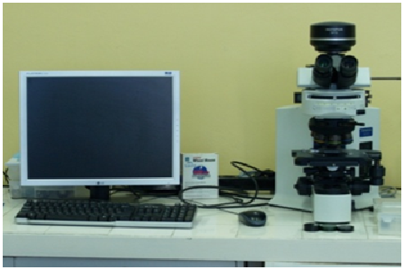
Microscope Olympus BX51
- UIS2 optical system
- Vertical stage movement: 25mm Motorized focus/vertical stage movement: 25mm, Stage stroke with coarse adjustment limit stopper 0.01µm increments, maximum speed: 3mm/s, Torque adjustment for coarse adjustment knobs coarse/fine changeover button, stage shunting button and Stage mounting position variable stage up/down button High sensitivity fine focusing knob (minimum adjustment gradations: 1µm)
- Motorized reflected fluorescence, 6-position mirror turret unit, motorized shutter changeover speed: shutter speed: 0.1 s
- 8-position with motorized AS, turret and top lens swing out mechanism (N.A. 1.4—0.9), for 1.25×* 1 * 2 —100×
- High sensitivity cooled CCD camera developed for live cell imaging
- New DIC observation system optimizes the specimen image at wider magnifications
- High-quality darkfield effect at all magnifications
- Polarizing observation for wide-area retardation measurement
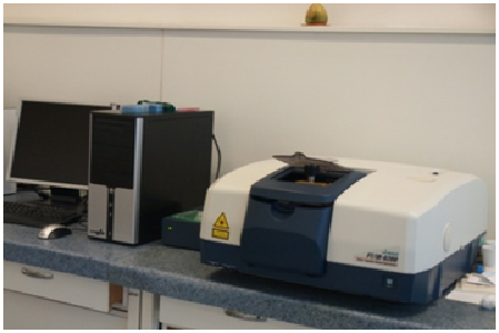
Spectrometer FT-IR JASCO 6200, with ATR Golden Gate
- Measurement wavenumber range: 7,800 to 350 cm-1
- Extended wavenumber range: 15,000 to 20 cm-1
- Display wavenumber range: 15,000 to 0 cm-1
- Wavenumber accuracy: Within ± 0.01 cm-1 (theoretical value)
- Resolution: 0.25 cm-1
- Sample chamber: Size: 200 mm (W) × 260 mm (D) × 185 mm (H) Optical path: Center focus, light axis 70 mm high
- Interferometer: 28° Michelson interferometer Corner cube mirror interferometer, with auto-alignment mechanism, sealed structure, DSP control
- Detector: DLATGS (with temperature regulator) (standard)
- Signal-to-noise ratio: 45,000:1
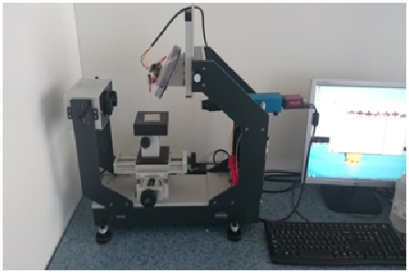
Drop Shape Analyzer DSA30, KRÜSS GmbH
- Measuring range for contact angle: 1-180°
- Measuring range for surface tension: 0.01-1000mN/m
- Measurement resolution for contact angle: 0.1°
- Measurement resolution for surface tension: 0.01mN/m
- Video system: CCD camera with IEEE1394b interface, 60fps, automatic triggering of measurement
- Optics: 6,5xzoom, FOV 3,5-23mm diagonal, integrated focus, high performance vario field illumination
- sample size: 300x-x50mm (WxDxH)
- Interfaces: RS232, IEEE1394b
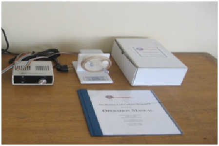
Rotative Cells Culture System, Synthecon
- The Rotary Cell Culture System (RCCS) is a unique 3D cell culture technology for culturing both suspension and anchorage-dependent cells. The RCCS-D bioreactor rotates one disposable vessel at a single speed; the rotation speed is adjustable.
- Includes: a single station rotator base, power supply, 4 disposable culture vessels.
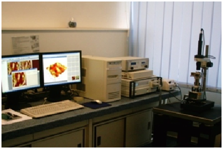
Atomic Force Microscope (Veeco Multi Mode VS-AM)
- characterization of biomaterials surface properties
- allows varying working conditions
- working Kit for magnetic field microscopy
- probe support for scanning in fluid
- command and control system for working with variable temperature
- in liquid working cell Kit for negative / positive temperature
- nanoindentation and scratching module
- A complete range of Atomic Force Microscopy (AFM) and Scanning Tunneling Microscopy (STM) techniques is available with the MultiMode SPM:
- Contact AFM— Measures topography by sliding the probe’s tip across the sample surface. Operates in both air and fluids.
- TappingMode™ AFM— Measures topography by tapping the surface with an oscillating tip. This eliminates shear forces which can damage soft samples and reduce image resolution. TappingMode is available in air and fluids.
- Non-contact AFM— Measures topography by sensing Van der Waals attractive forces between the surface and the probe tip held above the surface. Provides lower resolution than either contact AFM or TappingMode.
- Magnetic Force Microscope (MFM)— Measures magnetic force gradient distribution above the sample surface.
- Electric Force Microscope (EFM)— Measures electric field gradient distribution above sample surfaces.
- Surface Potential Microscopy— Measures differences in local surface potential across the sample surface.
- LiftMode™— A combined, two-pass technique that separately measures topography (using TappingMode) and another selected property (e.g., magnetic or electric force), using the topographical information to track the probe tip at a constant height above the surface.
- Force Modulation— Measures relative elasticity/stiffness of surface features. Force modulation is only one of several types of force imaging which are possible.
- Lateral Force Microscopy (LFM)— Measures frictional forces between the probe tip and sample surface.
- Scanning Tunneling Microscopy (STM)— Measures topography of surface electronic states using a tunneling current which is dependent on the separation between the probe tip and a conductive sample surface.
- Electrochemical Microscopy (ECSTM and ECAFM)— Measures the surface structure and properties of conducting materials immersed in electrolyte solutions with or without potential control.
- Lithography— Use of a probe tip to mechanically scribe or indent a sample surface. May be used to generate patterns, test surfaces for microhardness, etc.
- The unit is designed for imaging small (approx. 1.5 cm dia.) samples using a series of interchangeable scanners and is able to provide images from the atomic scale to 175 µm in size.
- Images consist of raster-scanned, electronic renderings of sample surfaces. There are three default sizes: 128 x 128 pixels, 256 x 256 pixels, and 512 x 512 pixels. In addition, six width-to-height aspect ratios may be specified by the user: 1:1, 2:1, 4:1, 8:1, 16:1, and 32:1.
- The MultiMode can scan up to 200 µm laterally (in X and Y) and 10 µm vertically (Z axis)
Teaching Activities
- Introduction in Biomaterials (1st year, graduate, Medical Engineering specialization)
- Biocompatibility (1st year, graduate, Medical Engineering specialization)
- Interaction biomaterials-tissue (2st year, master, Metallic Biomaterials program)
- Prosthetic explant analysis (2st year, master, Metallic Biomaterials program)
- Clinical Engineering (1st year, graduate, Medical Engineering specialization)
- Research Projects Management (1st year, graduate, Medical Engineering specialization)
- Surgical Instruments and Medical Devices (4st year, graduate, Medical Engineering specialization)
- Orthopedic implants and prosthesis (4st year, graduate, Medical Engineering specialization)
- Other: investigations for graduation and PhD thesis; Erasmus students from other countries.
Research Activities
- Interface Phenomena Implant-Tissue
- Surface Analysis
- Explant Analysis
- Biocompatible Coatings Characterization
- Biodegradable Metallic Biomaterials
- Biodegradation in Simulated Medium
- Bioceramics & Biocomposites
- Adhesion Phenomena
- Tissue Engineering
- Metallic Biomaterials
Training Activities
- Interface & Adhesion Phenomena in Biomaterials and Medical Devices
- Management of the Metallic Surgical Instruments
- Research Projects Management in Biomaterials and Medical Devices
Remarks
- Integration: Materials Characterisation (Materials Engineering field); Biomaterials & Medical Devices Characterization (Medical Engineering field).
- Complementarities: Sample Preparation Lab.; Quantitative light Microscopy Lab.; SEM/TEM Electron Microscopy Lab.; SDAR-OES & EDP XRF spectrometry Lab.; Biodegradation & Adhesion Lab.; Electrochemistry & Surface Functionalization Lab.; Corrosion Lab.